Ear Development
Inner Ear Development
Early in the 4th week, a thickening of the surface ectoderm on each side of the hindbrain -> otic placode -> otic pit -> form the otic (auditory) vesicle à membranous labyrinth. Each vesicle divides into:
The ventral component -> saccule and the cochlear duct (scala media)
The dorsal portion -> utricle, semicircular canals and endolymphatic duct and sac
Saccule, Cochlea, and Organ of corti
a. In the 6th week of development: saccular lower pole -> the cochlear duct -> penetrates surrounding mesenchyme in a spiral fashion -> completed 2.5 turns at the end of the 8th week. And the ductus reuniens connects the saccule with utricle.
b. Mesenchyme surrounding the cochlear duct -> forming cartilage.
c. In the 10th week, this cartilaginous shell undergoes vacuolization, and two perilymphatic spaces, the scala vestibuli and scala tympani.
d. The vestibular membrane separates cochlear duct from the scala vestibuli.
e. The basilar membrane separates cochlear duct from the scala tympani.
f. The spiral ligament attaches lateral wall of the cochlear duct to the surrounding cartilage.
g. The median angle of the cochlea duct is connected to and partly supported by a long cartilaginous process, the modiolus -> the future axis of the bony cochlea
h. The epithelial cells of the cochlear duct form two ridges:
i. The inner ridge, the future spiral limbus,
j. The outer ridge which forms the sensory hair cells a) *One inner row, b) *Three or four outer rows.
k. They are covered by the tectorial membrane.
l. The sensory cells + tectorial membrane ->constitute the organ of Corti.
m. Impulses received by this organ -> the spiral ganglion to -> the nervous system by the auditory fibers of 8th cranial nerve.
n. The capsular cartilage serves as a template for the later formation of the true bony labyrinth. The conversion from the cartilaginous to the bony labyrinth occurs between 16 and 23 weeks’ gestation
Utricle and Semicircular canals
a. During the 6th week of development, semicircular canals appear as flattened outpocketings of the utricular part of the otic vesicle -> Central portions of the walls of them -> appose each other and disappear à giving rise to 3 SCC. (Programmed cell death)
b. One end of each canal dilates -> form the crus ampullare, the other, the crus nonampullare, does not widen.
c. Five crura enter the utricle, three with an ampulla and two without.
d. Cells in the ampullae form a crest -> the crista ampullaris, containing sensory cells.
e. Similar sensory areas develop in the walls of the utricle and saccule.
f. Impulses generated in sensory cells of the cristae and maculae as a result of a change in body position à the brain by vestibular fibers of VIII cr n.
g. The statoacoustic ganglion forms during formation of the otic vesicle à The ganglion splits into cochlear and vestibular portions
Endolymphatic Duct and Sac
The auditory vesicle elongates and develops an indenting groove, which demarcates a tubular diverticulum on its medial side -> which will be modified into the endolymphatic sac and duct, -> and continues to grow postnatally until the age of three or four years
Pharyngeal arches
They are 5 arches developed by 23 days of gestation called mandibular arches in proximal portion of embryo, separated by pharyngeal grooves/clefts. They are 5 arches 1st, 2nd, 3rd, 4th and 6th. The 5th arch did not develop in human but in other species. Each arch is formed from:
Ectoderm: -> skin of head and neck region.
Endoderm: -> lining for oral, nasal, pharyngeal and Eustachian tube.
Mesoderm: core of each arch
Middle Ear development
The middle ear cavity and the auditory tube arise from -> first pharyngeal pouch called the tubotympanic sulcus, so -> lined with an endoderm.
This pouch expands in a lateral direction and comes in contact with the floor of the first pharyngeal cleft.
The distal part -> forming the tubotympanic recess.
The proximal part -> the auditory tube (Eustachian tube)
Later, the endodermal epithelium of the tubotympanic sulcus becomes close to the ectoderm lining the first pharyngeal cleft, separated by mesoderm. This complex forms the tympanic membrane (eardrum). During fetal life, a prominent ring-shaped, called the tympanic ring, supports the tympanic membrane. Later, the tympanic ring becomes absorbed into the temporal bone.
Just dorsal to the end of the tubotympanic sulcus, a condensation of mesenchyme -> appears at 6th weeks -> form the middle ear ossicles. The malleus and incus arise from mesenchyme of the 1st pharyngeal arch, whereas the stapes -> from 2nd arch. The tensor tympani muscle, which is attached to the malleus -> from first-arch mesoderm, so ->innervated by trigeminal nerve (cr n V). The stapedius muscle -> is of second-arch origin, and is innervated by the facial nerve (cr n VII).
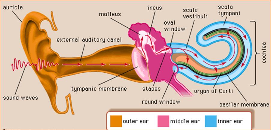
Ossicles -> lie in a bed of very loose embryonic connective tissue; extend from the inner layer of the tympanic membrane to the oval window of the inner ear. The future middle ear cavity remains filled with loose mesenchyme until late in pregnancy.
During the 8th and 9th months, programmed cell death and other resorptive processes -> clear the middle ear cavity and leave the auditory ossicles suspended within it. Free movement of the auditory ossicles is acquired within 2 months after birth.
During late fetal life, the tympanic cavity expands dorsally by vacuolization of surrounding tissue to form the tympanic antrum. And After birth, epithelium of the tympanic cavity invades bone of the developing mastoid process. Later, most of the mastoid air sacs come in contact with the antrum and tympanic cavity.
Development of the External Ear
The external auditory meatus à first pharyngeal cleft.
At the 3rd month, epithelial cells at the bottom of the meatus proliferate, forming a solid epithelial plate, the meatal plug.
In 7th month, this plug dissolves and the epithelial lining of the floor of the meatus participates in formation of the definitive eardrum.
Occasionally the meatal plug persists until birth, resulting in congenital deafness
Development of Auricle
By the end of 4th week, the auricle develops from 6 mesenchymal proliferations (hillocks) of 1st and 2nd pharyngeal arches, surrounding the first pharyngeal cleft.
These swellings (auricular hillocks), three on each side of the external meatus, later fuse and form the definitive auricle.
As fusion of the auricular hillocks is complicated, developmental abnormalities of the auricle are common.
Initially, the external ears are in the lower neck region, but with development of the mandible, they ascend to the side of the head at the level of the eyes.
Because of its association with the pharyngeal arches, the external ear is a sensitive indicator of abnormal development in the pharyngeal region. Other anomalies of the first and second arches are often attended by abnormally located external ears